

Ascending Aorta Aneurysm: Size, Monitoring and Surgery
Ascending aorta aneurysm surgery: learn size thresholds, monitoring protocols, and when intervention is needed.
What Is an Ascending Aorta Aneurysm?
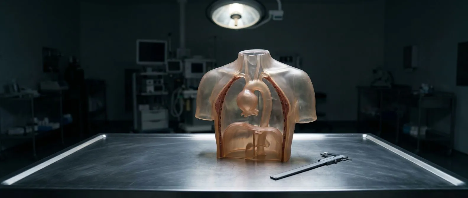
An ascending aorta aneurysm is a bulging or widening of the first upward-curving segment of the aorta, the large artery that carries oxygen-rich blood away from your heart. The ascending aorta begins at the left ventricle and arches upward toward the neck before curving back down through the chest and abdomen. When the muscular wall of this segment weakens, the pressure of each heartbeat pushes the vessel outward, much like a worn patch on an old inner tube. Over time, the diameter grows larger, and the wall grows thinner — increasing the risk of a tear (dissection) or rupture if left untreated.
Most patients discover an ascending aorta aneurysm by chance. The condition rarely produces symptoms until it becomes large enough to press on nearby structures. A chest X-ray ordered for a persistent cough, or an echocardiogram performed for an unrelated heart murmur, may reveal the dilated vessel before anything feels wrong. This incidental discovery is actually fortunate: it gives your cardiac team time to plan careful monitoring or, if the size crosses a guideline threshold, to schedule elective ascending aorta aneurysm surgery before an emergency occurs. According to Medical News Today, many thoracic aortic aneurysms are found during routine imaging for other conditions.
It helps to picture the ascending aorta as the first stretch of a major highway leaving the heart. The aortic valve sits at its base, opening with every contraction to release blood into the vessel. Just above the valve, the coronary arteries branch off, supplying the heart muscle itself. An aneurysm in this region can therefore affect valve function, coronary blood flow, and the structural integrity of the entire aortic root — which is why the decision to operate depends not only on diameter but on anatomy, growth rate, and your individual risk profile.
What Causes an Ascending Aorta Aneurysm?
The most common driver is long-standing high blood pressure, which subjects the aortic wall to relentless mechanical stress with every heartbeat. Over years and decades, the elastic fibres within the tunica media — the middle layer of the artery — fragment and thin, allowing the vessel to dilate. Atheromatous plaque, the fatty cholesterol deposit that narrows other arteries, can also weaken the aortic wall from within, disrupting the normal architecture and creating focal areas of stress. Patients with a family history of aortic disease face a substantially higher baseline risk.
Genetic and connective-tissue disorders are a second major category. Conditions such as Marfan syndrome, Loeys-Dietz syndrome, and vascular Ehlers-Danlos syndrome directly affect the proteins that give the aorta its tensile strength. In these patients, aneurysms can appear at a younger age and grow more quickly, sometimes reaching surgical thresholds in the thirties or forties rather than the typical sixth or seventh decade. If you have a first-degree relative who suffered an aortic dissection or sudden cardiac death, your surgeon will likely recommend genetic testing and baseline imaging regardless of symptoms. Elevated levels of apolipoprotein B, a cholesterol-carrying particle, have also been associated with faster aneurysm growth in some cohorts.
Other contributors include bicuspid aortic valve disease — a congenital valve defect present in roughly one to two percent of the population that alters blood flow patterns and stresses the adjacent aortic wall — as well as inflammatory conditions like giant cell arteritis and Takayasu arteritis. Previous heart surgery, chronic infections, and trauma can also leave the ascending aorta vulnerable. Understanding which mechanism is at work matters because it influences surveillance intervals: a Marfan patient may need annual scans, while a degenerative aneurysm measured at 4.2 cm might be safely monitored every two to three years.
How Is an Ascending Aorta Aneurysm Diagnosed?
Diagnosis combines imaging, physical examination, and a thorough family history. Your cardiologist will start with a transthoracic echocardiogram, which uses sound waves to visualise the aortic root and proximal ascending aorta through the chest wall. This painless study measures diameter in real time and also assesses valve function. If the echocardiogram suggests dilation, the next step is usually a computed tomography angiogram (CTA) or magnetic resonance angiogram (MRA), both of which provide highly detailed cross-sectional images of the entire aorta from the valve to the groin.
CTA versus MRA — which is better? Both are excellent, and the choice often depends on what else your team needs to learn. A CTA is faster and offers finer spatial resolution, making it the standard for surgical planning. An MRA avoids radiation, which matters for younger patients who will need lifelong serial imaging. Auscultation — listening with a stethoscope — may reveal a murmur from an associated valve problem, but it cannot measure aneurysm size. Only imaging can confirm diameter, and precise measurement matters: a difference of two millimetres can shift a patient from a watchful-waiting pathway to a recommendation for surgery. The Global Burden of Cardiovascular Diseases and Risk Factors, 1990–2019 underscores how underdiagnosis remains a global challenge, particularly in regions without routine access to advanced cardiac imaging.
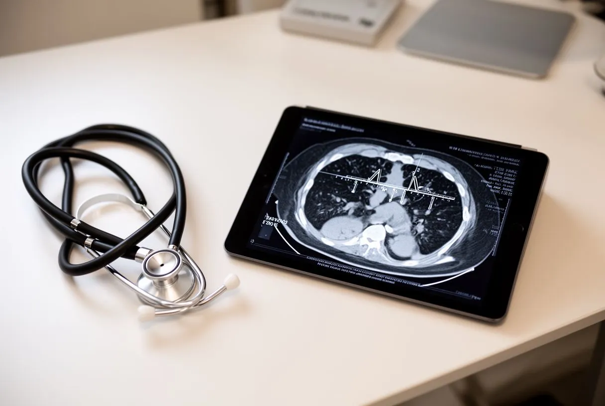
During your consultation, expect the surgeon to review your images with you on a screen, pointing out the aneurysm's maximal diameter, its relationship to the coronary arteries, and whether the aortic valve itself is involved. They will also ask about chest pain, back pain, shortness of breath, hoarseness, or a pulsating sensation in the upper chest — symptoms that, while uncommon with stable aneurysms, can signal rapid growth or impending complication. Bringing prior imaging to the appointment is invaluable: comparing measurements across years lets the team calculate growth rate, one of the most important factors in deciding whether and when to operate.
What Size Requires Ascending Aorta Aneurysm Surgery?
Size thresholds guide the conversation, but they are not rigid rules applied identically to every patient. For a degenerative ascending aorta aneurysm in a patient without genetic connective-tissue disease, the widely accepted surgical threshold is 5.5 cm. This number comes from population studies showing that the annual risk of dissection or rupture rises sharply above this diameter. The 2016 European Society of Cardiology guidelines, which remain a cornerstone reference for cardiac teams worldwide, endorse this 5.5 cm cut-off for elective intervention in standard-risk patients.
Lower thresholds for higher-risk patients apply in several situations. Patients with Marfan syndrome, Loeys-Dietz syndrome, or a family history of dissection at small diameters may be offered surgery at 4.5 to 5.0 cm. Those with a bicuspid aortic valve and an ascending aneurysm may cross into surgical territory at 5.0 cm, particularly if the valve itself needs repair or replacement. A rapid growth rate — defined as an increase of more than 3 mm per year on serial imaging — can also prompt earlier intervention regardless of absolute size. The underlying principle is that your surgical risk must be weighed against your rupture risk, and that balance shifts with each patient's anatomy, age, and overall health.
Your surgeon will also factor in EuroSCORE II, a validated calculator that estimates operative mortality based on variables such as age, renal function, lung disease, and urgency of the procedure. A patient with a low EuroSCORE II and a 5.3 cm aneurysm may reasonably be offered surgery earlier, because the procedure itself carries minimal added risk. Conversely, a patient with significant comorbidities and a 5.6 cm aneurysm might be counselled to continue surveillance if their operative risk is disproportionately high. The decision is always individualised, never dictated by the diameter alone.
How Does Ascending Aorta Aneurysm Surgery Work?
Ascending aorta aneurysm surgery is an open-chest procedure performed under general anaesthesia. The surgeon makes a vertical incision down the centre of the sternum (median sternotomy) to access the heart and great vessels. Once inside, the team establishes cardiopulmonary bypass — a heart-lung machine that takes over the work of pumping and oxygenating your blood so the heart can be still during the repair. An arterial cannula is placed, usually in the right axillary artery or directly in the aortic arch, to return oxygenated blood to your body while the ascending aorta is clamped and opened.
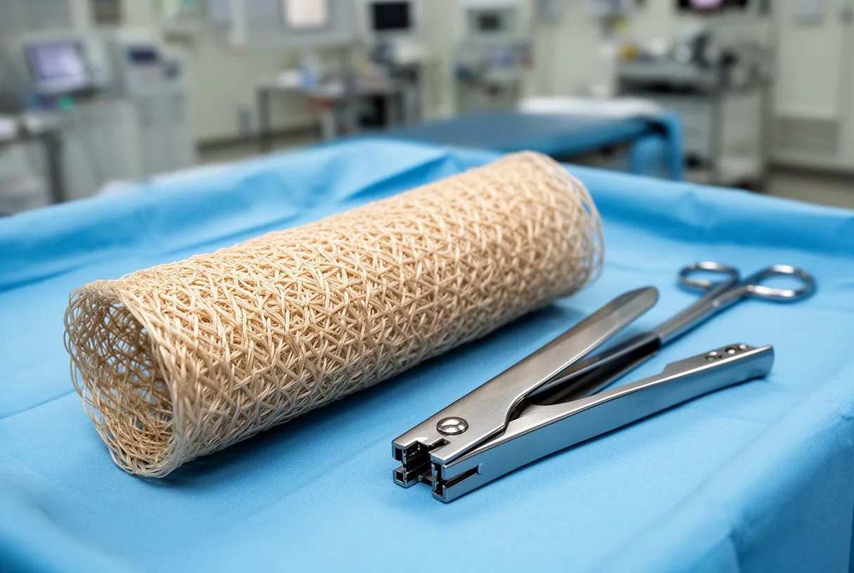
The diseased segment is resected and replaced with a synthetic graft made of woven polyester, a durable material that functions as a permanent new aorta. If the aneurysm involves the aortic root — the portion where the coronary arteries originate and the aortic valve sits — the surgeon may perform a Bentall procedure, replacing both the root and the valve and reimplanting the coronary arteries into the graft. When the native valve is healthy, a valve-sparing root replacement (the David or reimplantation procedure) can preserve your own valve while replacing the surrounding tissue. Autologous blood — your own blood, collected before bypass — is often used to prime the circuit and reduce reliance on donor transfusion.
The heart is then gradually weaned from the bypass machine, and the new graft is checked for leaks and proper flow. The sternum is closed with stainless-steel wires that remain in place permanently, and the skin is sutured or stapled. The entire operation typically takes four to six hours, depending on complexity. Throughout the procedure, the anaesthesia team monitors brain oxygenation and maintains a moderate hypothermia (cooling the body to around 28–32 °C) to protect the brain and other organs during the period when circulation is reduced.
What Are the Types of Ascending Aorta Aneurysm Surgery?
The specific operation depends on how much of the aorta is diseased and whether the aortic valve is involved. When the aneurysm is confined to the tube-like portion above the valve — sparing the root and the arch — a straightforward supracoronary graft replacement may suffice. The surgeon removes the aneurysmal segment and sews a polyester tube graft in its place, leaving the native valve and coronary ostia untouched. This is the least complex variant and generally carries the shortest bypass and cross-clamp times.
Root aneurysms require more complex reconstruction. The Bentall procedure replaces the aortic root, the valve, and the ascending tube in one integrated graft, reattaching the coronary arteries to the graft side. For patients whose valve is structurally normal, valve-sparing operations reconstruct the root around the existing valve leaflets, avoiding the lifelong need for blood thinners that comes with a mechanical valve. If the aneurysm extends into the aortic arch — the curved portion that gives off the head and neck vessels — a hemiarch or total arch replacement may be performed, sometimes with a frozen-elephant-trunk technique that extends a stent graft down into the descending aorta to address downstream disease in a single stage. An arterial graft harvested from elsewhere in the body may be used in specific reconstruction scenarios.
Valve choice, when valve replacement is necessary, involves a detailed conversation. Mechanical valves last indefinitely but require lifelong anticoagulation with warfarin. Biological tissue valves — made from cow or pig pericardium — typically do not require long-term blood thinners but may wear out after fifteen to twenty years. The decision considers your age, lifestyle, tolerance for anticoagulation monitoring, and future pregnancy plans. There is no universally right answer; the goal is to find the lowest-risk option for your situation.
What to Expect During Ascending Aorta Aneurysm Surgery Recovery
Recovery from ascending aorta aneurysm surgery unfolds in stages. The first twenty-four to forty-eight hours are spent in the intensive care unit, where nurses and doctors monitor heart rhythm, blood pressure, drainage from chest tubes, and neurological status. You will likely be breathing with the help of a ventilator for the first few hours after surgery, with the tube removed once you are fully awake and breathing independently. Most patients sit up in a chair on the first postoperative day and begin short walks with assistance by day two or three.

The hospital stay averages five to seven days. Pain around the sternotomy incision is expected and is managed with a combination of intravenous and oral analgesia, tapered steadily as healing progresses. You may notice a dry cough from the breathing tube and mild swelling in your legs from fluid shifts — both resolve within the first week. Low-dose aspirin is typically started to reduce the risk of clot formation on the graft or any valve component. If you received a mechanical valve, warfarin dosing begins in the hospital and continues indefinitely, with regular INR checks. Conditions such as atrial flutter, an irregular heart rhythm that can surface after cardiac surgery, are usually transient and managed with medication or a brief course of cardioversion.
At home, full recovery takes roughly eight to twelve weeks. You will be advised to avoid lifting anything heavier than five kilograms, refrain from driving for four to six weeks, and keep the incision clean and dry. A structured cardiac rehabilitation programme — supervised exercise, education, and lifestyle counselling — typically begins four to six weeks after discharge and continues for several months. Arm pain, especially in the left shoulder, is common as the nerves around the sternum recover; gentle range-of-motion exercises help. Most patients return to office work in six to eight weeks and to all normal activities, including exercise, by three months — though your surgeon will clear you based on your individual progress, not a fixed calendar.
Are There Alternatives to Ascending Aorta Aneurysm Surgery?
For aneurysms below the surgical threshold, active surveillance is the primary alternative and is not simply doing nothing. It involves serial imaging at defined intervals — typically every one to two years for moderate-sized aneurysms — to track diameter and growth rate. Blood pressure control is essential: your cardiologist will target a systolic pressure below 130 mmHg, often using beta-blockers or angiotensin receptor blockers, which reduce the sheer force exerted on the aortic wall with each heartbeat. Statins may be prescribed to slow atherosclerotic progression, and smoking cessation is non-negotiable.
Endovascular approaches have expanded dramatically in recent years, though they remain less common for the ascending segment than for the descending aorta. Devices are being developed for endovascular ascending repair, but these are not yet standard practice for most patients and are typically reserved for those too frail for open surgery in specialised centres. Balloon angioplasty and balloon aortic valvuloplasty are techniques used for valve disease, not for aneurysm repair; they cannot reduce the diameter of a dilated aorta. Transcatheter aortic valve replacement (TAVR), which uses a balloon-expandable valve deployed through a catheter, addresses valve stenosis but does not replace the aneurysmal aortic wall tissue.
In rare cases, an awake ECMO (extracorporeal membrane oxygenation) strategy may be used as a bridge in emergency situations, but this is a temporary support measure rather than a treatment for the aneurysm itself. The honest answer is that, once an ascending aorta aneurysm reaches the size where rupture risk outweighs surgical risk, open repair remains the definitive and gold-standard intervention. Medical therapy can slow growth, but it cannot reverse an established aneurysm.
What Are the Risks of Ascending Aorta Aneurysm Surgery?
Modern ascending aorta aneurysm surgery is performed with a high degree of safety in experienced centres, but it remains major surgery and carries real risks. Operative mortality for elective procedures in low-risk patients is generally reported at one to three percent, rising substantially for emergency operations performed after dissection or rupture. Bleeding requiring reoperation, infection of the sternum, stroke, kidney injury, and prolonged ventilation are among the most significant complications. Your surgeon will discuss your individual EuroSCORE II estimate, which predicts operative risk based on your specific health profile.
Neurological risk deserves particular attention because the ascending aorta and arch supply blood to the brain. Stroke rates range from one to four percent in elective cases, and techniques such as selective cerebral perfusion and hypothermic circulatory arrest are used to protect the brain during the period when the aorta is open. Atypical infarction — a heart attack that presents without classic chest pain — can occur in the perioperative period due to coronary reimplantation or air emboli. The anaesthesia and surgical teams use transoesophageal echocardiography and continuous monitoring to detect problems in real time. If a dangerous arrhythmia develops, the team is prepared to use an automated external defibrillator or intravenous antiarrhythmic medication immediately.
Long-term considerations include graft durability — polyester grafts are designed to last a lifetime and rarely require revision — and valve-related issues if a biological valve was implanted. Patients with mechanical valves must manage anticoagulation carefully, as both under-treatment (stroke risk) and over-treatment (bleeding risk) carry serious consequences. The ARISTOTLE trial and similar studies have shaped contemporary dosing guidance for apixaban in atrial fibrillation, but DOACs are not used with mechanical heart valves, which require warfarin or another vitamin K antagonist in both the aortic and mitral positions. Regular follow-up imaging — typically a CT scan or MRI at one year, then every two to three years — ensures the graft and any remaining aortic segments remain stable over time.
When Should You Seek Help for an Ascending Aorta Aneurysm?
If you are under surveillance for a known aneurysm, certain symptoms demand immediate emergency evaluation regardless of your scheduled imaging date. Sudden, severe chest or back pain — often described as tearing or ripping — is the hallmark sign of acute aortic dissection and constitutes a life-threatening emergency. Call emergency services or go to the nearest emergency department immediately. Other warning signs include new or worsening shortness of breath, difficulty swallowing, hoarseness, a pulsating sensation in the chest, or any neurological change such as weakness, numbness, or visual disturbance.
Routine follow-up is equally important even when you feel perfectly well. Aneurysms grow silently, and the only way to know whether you have crossed a surgical threshold is to keep your imaging appointments. If your last scan was more than two years ago and you have lost contact with your cardiac team, schedule a follow-up. Bringing previous imaging to any new consultation allows the surgeon to calculate growth rate, which is critical information that no single scan can provide. For patients with genetic syndromes or family history, first-degree relatives should also undergo baseline screening — an echocardiogram and, if indicated, a CT or MRI — to rule out inherited aortic disease.
- Annual check-ups: Even after successful surgery, yearly cardiac reviews and imaging every two to three years are standard.
- Know your number: Keep a record of your aneurysm diameter from each scan so you can track growth over time.
- Red-flag symptoms: Tearing chest or back pain, sudden weakness, fainting, or a new pulsating mass in the chest require emergency care.
- Family screening: First-degree relatives of any patient with an ascending aorta aneurysm should undergo baseline aortic imaging.
This guide provides general information to help you understand the anatomy, the decision-making framework, and what to expect before and after ascending aorta aneurysm surgery. It does not replace a personal consultation. Every aneurysm, every heart, and every patient is different. If you or a loved one has been diagnosed with an ascending aorta aneurysm — or if you have a family history that concerns you — the most important step is to discuss your individual situation with a qualified cardiac surgical team. Dr Alejandro Molina and the team at Cirugía de Corazón, with practices in Guadalajara, Querétaro, and Hermosillo, are available to review your imaging, explain your options in plain language, and help you decide the safest path forward. Book a consultation rather than self-diagnosing — the conversation itself may be the most reassuring step you take.






