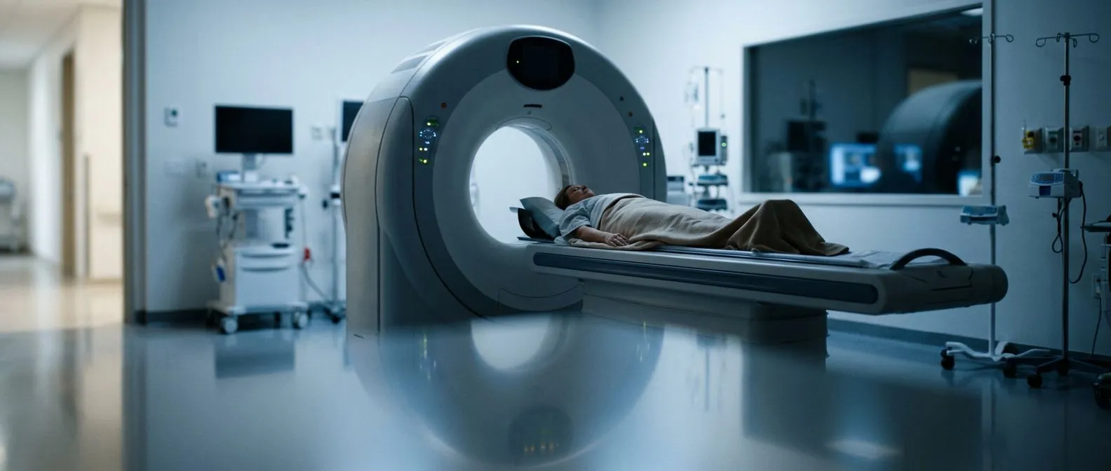
Surveillance Imaging After Aortic Surgery
Surveillance imaging after aortic surgery helps catch complications early.
Surveillance imaging after aortic surgery is the planned series of scans that checks how your aorta and your repair are holding up over the years. If you or a family member has had an aneurysm repaired, a dissection treated, or a valve procedure on the aortic root, these scans are not an optional extra — they are part of the treatment itself. This guide walks you through which tests are used, how often they happen, what each one shows, and what you should expect at every step, so the whole process feels less mysterious and far less frightening.
What Is Surveillance Imaging After Aortic Surgery?
Surveillance imaging after aortic surgery simply means taking pictures of your aorta at planned intervals after an operation, to confirm the repair is stable and to catch any change early. The aorta is the body's largest artery, running from the heart down through the chest and abdomen. Surgery on it — whether open repair with a graft or a minimally invasive stent graft — creates a new set of structures that need watching over time.
The logic is much like the check-ups you have after dental work or after placing a stent in a coronary artery: the problem was fixed once, but the vessel lives with you for the rest of your life. Blood pressure, aging, and the underlying disease that caused the original problem can all slowly change the aorta again. Imaging finds those changes while they are still small and manageable.
A written imaging plan helps, because a follow-up schedule you can actually see is one you are more likely to follow.
Why Do You Need Follow-Up Imaging After Aortic Surgery?
The aorta can change even after a successful operation. A graft itself is strong, but the aorta above and below it remains living tissue. New aneurysms can form in segments that were once normal, and the connection points where the graft meets the vessel — called anastomoses — can weaken or dilate. After a Type A Aortic Dissection repair, the remaining dissected aorta needs lifelong observation because the false channel may enlarge over the years.
Early detection changes what treatment you need. A small change found on a routine scan might mean adjusting blood pressure medication or simply shortening the interval to the next scan. The same change discovered years later, after symptoms appear, might mean emergency surgery. Guidelines from major cardiovascular societies consistently recommend structured follow-up for precisely this reason — problems found on scheduled scans are almost always easier and safer to treat than problems found in the emergency room.
This is also true after procedures on the aortic valve. Whether you had a Surgical Aortic Valve Replacement or a Transcatheter Aortic Valve Implantation, the aortic root and the valve itself need periodic checking for function, position, and any widening around the new valve. If you are considering any of these procedures and want a second opinion on the follow-up plan that comes with them, that is a normal and sensible request — we encourage it. Our multidisciplinary team reviews every case against international guidelines before recommending a procedure, and we present the alternatives along with the follow-up burden each option carries.
What Is Digital Surveillance in Heart Care?
Digital surveillance is a term you may see in the search results, and it means something different from medical follow-up. In technology and security, digital surveillance refers to monitoring of data, networks, or activity. Some hospitals also use the phrase for remote monitoring of implanted devices, such as pacemakers that transmit readings to the clinic automatically. It is not the same thing as the CT and MRI scans covered in this article.
It is worth clarifying because search engines mix the two meanings freely. When you search for "surveillance imaging," you may land on pages about security cameras or data monitoring. In the context of aortic surgery, surveillance always means periodic medical imaging of the blood vessel itself — performed with scanners, interpreted by radiologists and cardiac Imaging on Disc records, and reviewed by your surgical team.
The closest heart-related cousin of digital surveillance is remote device follow-up. If you have a pacemaker or an implanted loop recorder, its readings can often be checked from home. Your aorta, however, still needs the scanner — there is no remote way to measure an aneurysm sac or a graft endoleak.
What Are the 7 Types of Imaging Used After Aortic Surgery?
Imaging comes in several forms, and each answers a different question. When people ask "what are the 7 types of imaging?", they usually mean the major modalities used in medicine. Here they are, with what each one does for aortic patients:
- X-ray: quick, low-dose images, mainly used for chest and lung checks rather than aortic detail.
- CT scan (computed tomography): the workhorse of aortic follow-up; gives detailed cross-sectional images of the graft, the aneurysm sac, and the whole vessel.
- MRI (magnetic resonance imaging): detailed imaging without ionising radiation; useful in younger patients who need many decades of scans.
- Echocardiogram: ultrasound of the heart; checks valve function and the aortic root.
- Vascular ultrasound: painless sound-wave pictures of the abdominal aorta, often used for simple surveillance of an Abdominal Aortic Aneurysm.
- Nuclear imaging: shows blood flow and inflammation; occasionally used when infection of a graft is suspected.
- Angiography (catheter-based): invasive imaging with dye injected through a thin tube; now mostly reserved for cases where treatment is planned in the same sitting.
You will not need all seven. Most aortic patients cycle between CT, echocardiography, and sometimes MRI. Is radiology considered a doctor? Yes — the specialist who reads your scans is a radiologist, a fully qualified physician, and their report is what your surgeon works from. As for what medical imaging job pays the most, that is a career question rather than a patient one, but it does reflect how much training goes into reading these images correctly — expertise you want on your side.
CT Scan After Aortic Surgery: What to Expect
The CT scan is the standard test for aortic surgery follow up imaging. A CT scanner is a large doughnut-shaped machine; you lie on a table that slides through it while X-ray images are taken in seconds. For aortic studies, iodine dye is injected into a vein so the vessel and the graft light up clearly. Modern protocols keep the radiation dose as low as possible while still producing diagnostic pictures.
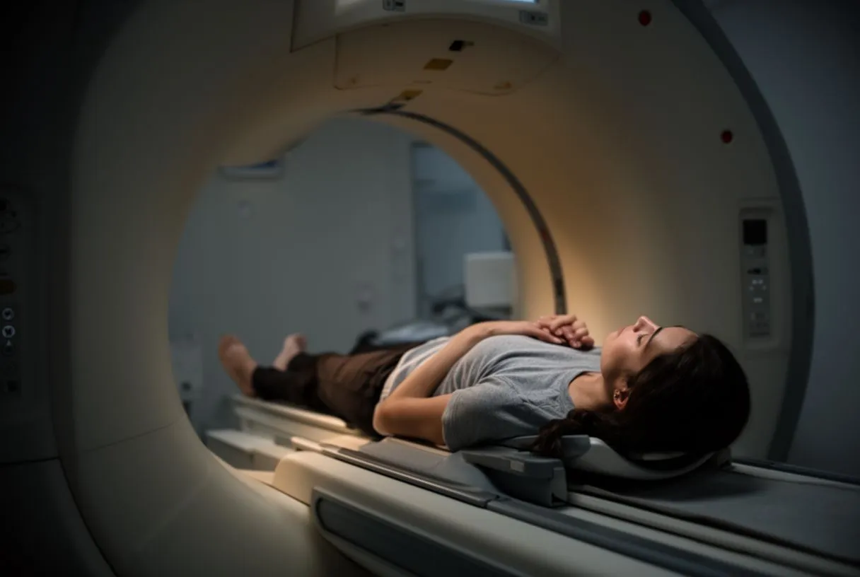
What the radiologist measures matters more than the scan itself. The team compares every study with your previous ones: total Aortic Diameter at set levels, the size of any residual false channel after dissection, graft position, and blood flow outside the graft (called an endoleak after stent-graft repair). Standardised measurement protocols matter enormously here — a consensus document by the European Society of Cardiovascular Radiology lays out exactly how CT and MR scans should be performed and reported before procedures such as valve implantation, so that measurements taken in different hospitals can be compared reliably (European Society of Cardiovascular Radiology consensus document).
Preparation is simple, and the scan is painless. You may be asked to fast for a few hours and to have a blood test to check kidney function before the dye is given. The whole visit usually takes under an hour. In our consultation rooms, we take the time to explain what the studies actually show, in plain words, before any decision is made — bring your questions, and expect them to be answered. We always review the images together with the patient rather than just quoting the radiology report, because seeing your own aorta on the screen makes the plan real.
Echocardiogram After Aortic Surgery: Why It Matters
An echocardiogram is an ultrasound of your heart, and it is usually the first test at every follow-up visit. It is painless, involves no radiation and no dye, and takes about twenty minutes. For patients who have had a valve procedure — whether open or transcatheter — the echocardiogram tells the team how the new valve is opening and closing, whether it is leaking, and how well the heart muscle is working.
It also watches the aortic root and the Aortic Annulus. The annulus is the ring of tissue where the valve sits, and its size matters for valve choice and for future procedures. After an aortic root replacement, echocardiography tracks the graft and the valve function together, while CT tracks the wider aorta. The two tests complement each other rather than compete.
Screening programmes are increasingly finding Severe Aortic Stenosis early in the community — one example is a study that added echocardiography to routine flu vaccination visits to detect valve disease before symptoms appear (ClinicalTrials.gov). The same principle applies after your surgery: regular, simple checks find problems while they are still easy ones.
MRI Surveillance After Aortic Repair: When Is It Used?
Cardiac MRI after aortic root replacement and other repairs is chosen when avoiding radiation matters. MRI uses a powerful magnetic field and radio waves — no X-rays at all. For younger patients facing decades of annual scans, or for those with kidney problems who cannot safely receive iodine dye, MRI is often the better long-term choice. It gives excellent pictures of the aortic wall, the graft, and blood flow through the vessel.
MRI does have practical trade-offs. The scanner is loud, the appointment is longer (45 to 90 minutes), and some implanted devices are not MRI-compatible — though most modern heart devices are. Claustrophobia is real for some patients; tell your team beforehand, as mild sedation is an option. The same European standardisation document mentioned above covers how MRI protocols should be structured so that every scan in your series is comparable with the last.
In practice, the choice between CT and MRI is individual. It depends on your age, your kidney function, the type of repair, the segment of aorta involved — including the Aortic Arch, which is harder to image than the descending vessel — and how frequently you need scans. This is a conversation, not a default.
What Is the Aortic Aneurysm Monitoring Schedule?
The schedule depends on the size of the aneurysm and the type of repair you had. Before surgery, aneurysms are monitored with imaging at intervals set by their diameter; guideline thresholds such as an aortic diameter above 5.5 cm generally prompt consideration of repair, earlier in connective tissue conditions like Marfan syndrome. After repair, surveillance continues — because new aneurysms can form elsewhere in the aorta.
A typical pattern after open repair looks like this:
- Before discharge: a baseline scan confirming the repair is sound.
- Around one month: an early check of the healing connection points.
- Every 6 to 12 months for the first years: CT or MRI of the repaired segment and the rest of the aorta.
- Annually once things are stable: with echocardiography added as needed.
- Immediately, at any time: new chest, back, or abdominal pain always triggers urgent imaging, whatever the calendar says.
Your underlying condition shapes the plan. Patients with a Thoracic Aortic Aneurysm from a genetic condition need closer and longer surveillance than someone with an isolated degenerative aneurysm. After repair of an Acute Aortic Syndrome — the umbrella term for dissection, intramural haematoma, and Penetrating Aortic Ulcer — the residual aorta is monitored closely, because disease can progress in segments that were not operated on. Abdominal Aortic Aneurysm follow-up, whether repaired open or by stent graft, follows its own well-established rhythm of ultrasound and CT checks.
TEVAR Follow-Up Imaging Frequency for Endovascular Repair
Endovascular repair — placing a stent graft inside the aorta through small incisions in the groin — needs the most diligent imaging of all. TEVAR (thoracic endovascular aortic repair) and its abdominal equivalent, EVAR, depend on the stent graft staying sealed against the vessel wall and in the right position. Over time, the graft can shift, the seal can leak (an endoleak), or the aneurysm sac can fail to shrink the way it should.
The typical endovascular aortic repair surveillance timeline runs: a scan within the first month, another around six to twelve months, and then annually — usually CT with contrast, sometimes supplemented by ultrasound. Each scan checks graft position, endoleak presence, sac size, and changes at the landing zones where the graft grips the healthy vessel. Missing a year matters: endoleaks can be silent and still require treatment.
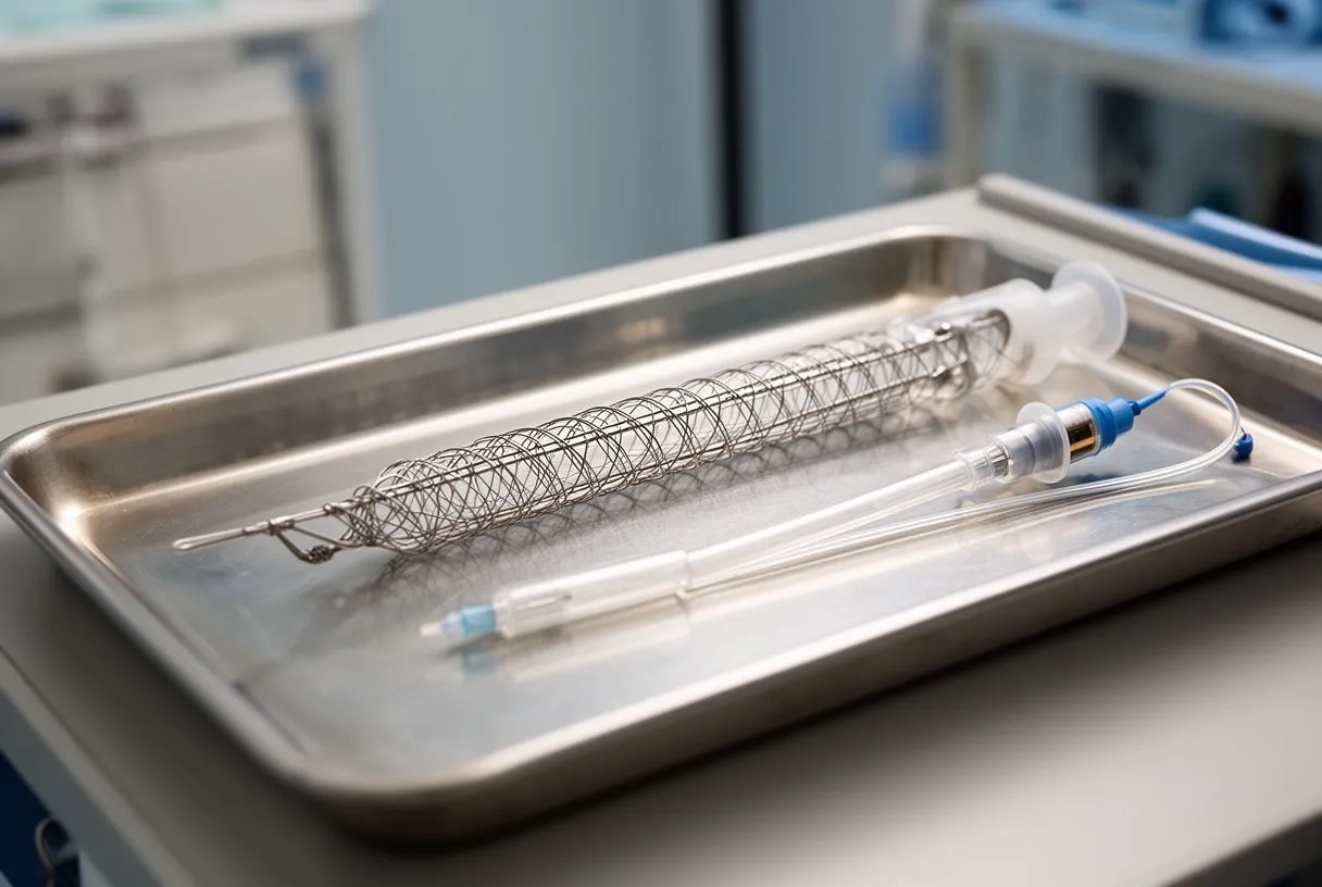
This is why we tell every stent-graft patient that the procedure is only half the treatment — the other half is the imaging calendar. Scheduling the first follow-up scan before leaving the hospital means surveillance starts on day one rather than whenever someone remembers.
When to Repeat Imaging After Aortic Surgery — and When to Call Your Team
Follow the calendar, but also trust your symptoms. The scheduled scans catch slow changes. Sudden symptoms catch nobody's schedule and need immediate attention. Call emergency services — do not wait for a routine appointment — if you experience any of the following:
- Sudden severe chest, back, or abdominal pain — especially pain that is tearing or moves position, which can signal Aortic dissection: a true cardiovascular emergency, whether Type A Aortic Dissection or Type B Aortic Dissection.
- Fainting, sudden weakness, or loss of pulse in an arm or leg.
- Shortness of breath that comes on abruptly, or symptoms of shock.
Routine changes also deserve a call, without panic. A new murmur heard by your family doctor, new breathlessness climbing stairs, or an abnormal finding on a scan done elsewhere — these belong in front of your aortic team promptly, but calmly. If you have an Intra-aortic Balloon Pump or other temporary support device during a hospital stay, your team will arrange imaging as part of that care; it does not change your long-term plan.
Choosing where your follow-up happens matters as much as having it. Consistency of technique and reporting is what makes scans comparable year to year, and the best hospital for aortic surgery follow-up imaging is one that keeps your full image archive, uses standardised protocols, and has a multidisciplinary team reading the results. Whether your repair was an Aortic Aneurysm Repair, an Aortic Arch Replacement, or a valve-sparing root procedure, ask whoever treats you: what will we look at, when, and what would change the plan? Those three questions turn surveillance from a worry into a tool.
Surveillance imaging after aortic surgery is not a footnote to your operation — it is the second half of the treatment that keeps the first half working. Know your schedule, keep every scan in one place, compare each result with the last, and never let new symptoms wait for the calendar. If you are unsure whether your current follow-up plan is complete, or you would like a second opinion on your aortic repair and its monitoring, our team offers a free initial video consultation where we review your history, explain your studies in plain language, and tell you which scans are genuinely needed. Book a consultation with a qualified cardiac team rather than second-guessing on your own — your aorta, and your peace of mind, deserve the professional version of vigilance.