

Aortic Rupture: Warning Signs and Emergency Repair
Aortic rupture warning signs can save your life—learn the tearing chest pain signals, emergency diagnosis, and repair options at our Guadalajara cardiac center.
When the body's main pipeline for oxygen-rich blood fails, every minute matters. Recognizing aortic rupture warning signs early can mean the difference between a treatable condition and a life-threatening emergency. For patients and families facing the anxiety of cardiac disease, understanding what happens inside the chest—without drowning in medical jargon—provides a crucial foundation for making calm, informed decisions. This guide bridges complex cardiovascular surgery with clear, empathetic explanations, focusing on what you experience, how the anatomy responds, and what repair entails.
What Are Aortic Rupture Warning Signs
Sudden, tearing chest or back pain: The most classic of all aortic rupture warning signs is a severe, ripping sensation that travels from the chest down through the back. Unlike the heavy, crushing pressure of a standard heart attack, this pain feels sharp and migratory, reflecting the physical tearing of the vessel wall. Patients often describe it as the worst pain they have ever felt, arriving without warning and peaking in intensity within moments. Recognizing this specific pain profile is critical for emergency responders.
Pulse or blood pressure discrepancies: Because the aorta branches to supply the arms and legs, a tear can partially block these pathways. A physician checking your vitals might find a significantly different blood pressure reading in your left arm compared to your right arm. Similarly, one pulse might be weak or absent entirely while others remain strong. These asymmetrical vital signs are massive red flags pointing directly toward aortic compromise.
Neurological symptoms and fainting: If the dissection or rupture affects the arteries supplying the brain, patients may experience sudden dizziness, difficulty speaking, weakness on one side of the body, or a temporary loss of consciousness. This occurs because the torn aortic flap is obstructing blood flow to the carotid arteries. Any sudden fainting episode accompanied by severe chest pain warrants immediate emergency medical attention.
How the Ascending Aorta Fails and Tears
The aorta is the largest artery in the human body, originating directly from the left ventricle of the heart. The first segment, known as the ascending aorta, curves upward and carries the entire cardiac output before branching off to the rest of the body. This segment experiences the highest direct pressure from the heart's pumping action. Over years of uncontrolled hypertension, this pressure stresses the elastic tissues of the vessel wall, causing them to stiffen and weaken.
When the inner lining of this vessel tears, blood surges through the gap, splitting the middle and outer layers of the arterial wall. This process is called a dissection. As the false channel fills with blood, it can eventually burst outward, causing a true aortic rupture. Accumulation of atheromatous plaque inside the artery can further weaken these walls, creating localized points of structural failure where the tissue is thinnest and most vulnerable to tearing.
Genetics also play a profound role in the structural integrity of this vessel. Conditions like Marfan syndrome, Loeys-Dietz syndrome, or vascular Ehlers-Danlos syndrome produce defective connective tissues from birth. In these patients, the aortic wall is inherently fragile and prone to stretching into an aneurysm long before high blood pressure ever becomes a factor. Monitoring the aortic diameter using echocardiograms is essential, as surgical guidelines recommend elective repair once the diameter exceeds 5.5 cm to prevent emergency ruptures.
When Chest Pain Signals an Emergency
Distinguishing from a typical heart attack: A myocardial infarction occurs when a coronary artery is blocked, starving the heart muscle of oxygen. While both produce severe chest pain, aortic rupture warning signs include that specific radiating, tearing quality. Occasionally, a dissection can shear the coronary arteries, causing an atypical infarction where both conditions occur simultaneously, making rapid hospital evaluation crucial.
Radiation beyond the chest: While the pain originates centrally, it frequently radiates down the spine, into the abdomen, or even into the neck and jaw. Some patients also report sudden, severe arm pain if the subclavian arteries are compromised by the tearing process. The broad, shifting nature of the discomfort is a distinguishing clinical feature that helps physicians differentiate aortic emergencies from standard cardiac events.

Immediate actions to take: If you or a loved one experience sudden, severe, tearing chest pain, do not attempt to drive to the hospital. Call your local emergency number immediately. Paramedics are equipped to monitor vital signs in both arms, begin intravenous blood pressure management to reduce the force against the aortic wall, and route you to a facility capable of rapid CT imaging and emergency cardiac surgery. Time is tissue, and in this case, time is life.
Understanding Acute Aortic Syndromes
Acute aortic syndrome is an umbrella term used by cardiologists to describe a group of life-threatening aortic emergencies. It includes aortic dissection, intramural hematoma (blood pooling within the wall without a tear in the lining), and penetrating atherosclerotic ulcer. All three present similarly and require urgent imaging to differentiate. During an initial clinical exam, careful auscultation with a stethoscope might reveal a new heart murmur, suggesting the aortic valve is being damaged by the expanding hematoma.
Dissections are categorized by where they begin. Type A involves the ascending aorta and is an immediate surgical emergency requiring open repair to prevent death from cardiac tamponade or stroke. Type B originates in the descending aorta and is often managed medically or with endovascular stenting, though emergency surgery is required if it ruptures. Understanding these classifications helps families grasp why two patients with similar symptoms might receive vastly different treatments.
- Type A Dissection: Involves the ascending aorta; requires immediate open surgery to save the heart and brain.
- Type B Dissection: Involves the descending aorta; often managed medically with blood pressure control unless rupture occurs.
- Intramural Hematoma: A bruise within the aortic wall that can mimic dissection; monitored closely for progression.
How Aortic Rupture Is Diagnosed Quickly
Upon arrival at the emergency department, rapid confirmation of aortic rupture warning signs is paramount. The gold standard for diagnosis is a computed tomography (CT) angiogram. This imaging uses a contrast dye injected into a vein to illuminate the aorta in three dimensions, revealing exactly where the tear is, how far it extends, and which major branches are compromised. This scan dictates the entire surgical strategy.
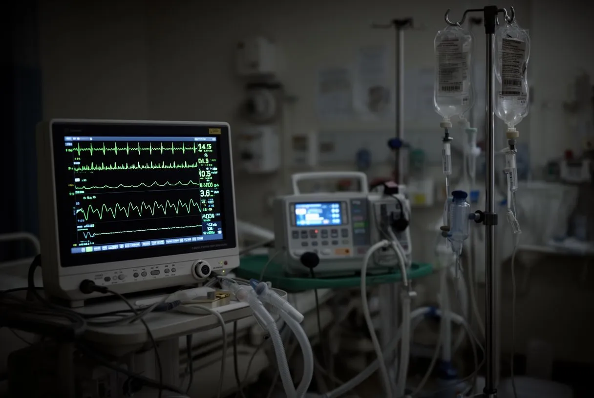
Because blood pressure management is critical to prevent the tear from extending, an arterial line is usually inserted into the wrist immediately. This invasive catheter provides beat-by-beat blood pressure readings, allowing critical care teams to titrate medications precisely. Intravenous beta-blockers and vasodilators are administered to lower the heart rate and blood pressure, reducing the sheer force pounding against the fragile aortic wall.
For patients who cannot undergo a CT scan, a transesophageal echocardiogram (TEE) provides an alternative view. By placing an ultrasound probe down the esophagus, physicians get a remarkably clear image of the ascending aorta and the heart valves. Rapid, accurate imaging is the cornerstone of survival, as delays in diagnosis increase the mortality rate significantly with every passing hour. Trusted medical resources like Verywell Health emphasize that prompt imaging is the linchpin of aortic emergency care.
Emergency Repair Options for Aortic Tears
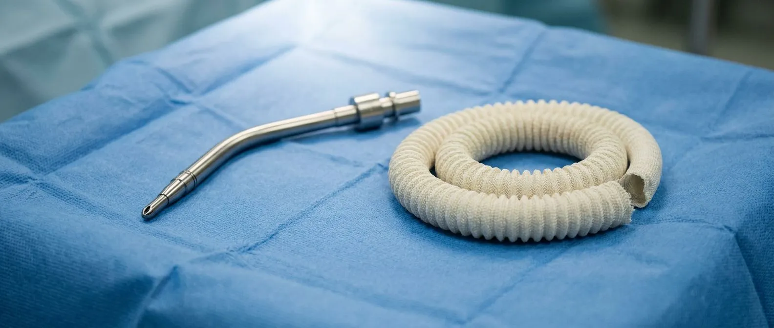
For Type A dissections and ruptures of the ascending aorta, open surgical repair is mandatory. The cardiac surgeon must open the chest, locate the tear, and replace the damaged segment of the aorta with a synthetic arterial graft. This is a massive operation that often requires stopping the heart and using a cardiopulmonary bypass machine to maintain circulation to the brain and body while the delicate suturing takes place.
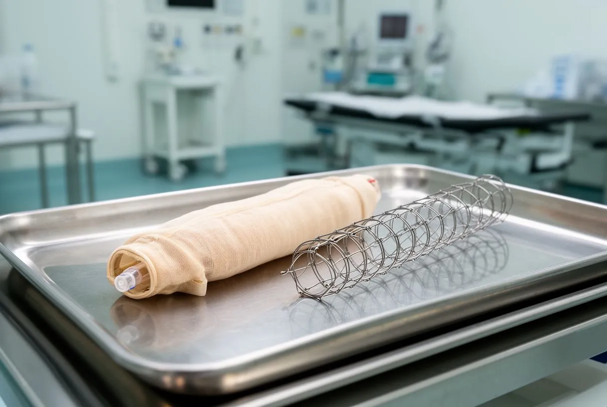
Valve-sparing vs replacement techniques: Often, the tear damages the aortic valve, causing it to leak. Surgeons may attempt to repair the native valve, or they might replace it using a mechanical or biological substitute. In some pediatric or highly specific adult cases, an autograft (the patient's own tissue) might be used, though synthetic polyester grafts are the standard for adults. The goal is to restore a stable, durable architecture to the heart's outflow tract.
For Type B emergencies, endovascular repair has revolutionized treatment. Instead of open surgery, a catheter is threaded through the femoral artery in the groin, guiding a stent graft up into the descending aorta. This stent expands to seal the tear and redirect blood flow through a reinforced channel. This minimally invasive approach dramatically reduces the physical trauma of surgery and is often the safest option for patients with multiple comorbidities. Research on complex cardiovascular emergencies, such as the Occurrence of Fatal and Nonfatal Adverse Outcomes after Heart Transplantation in Patients with Pretransplant Noncytotoxic HLA Antibodies, highlights the severe stakes involved when complex surgical interventions are delayed.
Recovery Timeline After Emergency Aortic Surgery
The first few days following emergency aortic repair are spent in the intensive care unit (ICU). Patients are heavily monitored for bleeding, neurological changes, and organ function. Blood pressure is meticulously controlled to protect the fragile new graft. To minimize the need for donor transfusions, modern surgical teams utilize cell salvage techniques, returning the patient's own autologous blood to their circulation to maintain volume without triggering immune responses.
Recovery from a Type A open repair is arduous. Patients typically spend a week or more in the hospital, progressing from bed rest to sitting, and eventually walking with assistance. Pain management, pulmonary therapy to prevent pneumonia, and gradual cardiac rehabilitation are the pillars of this phase. If the lungs are struggling to oxygenate blood, advanced support like awake ECMO (Extracorporeal Membrane Oxygenation) might be employed, allowing the patient to remain conscious while the machine bypasses their heart and lungs.
Long-term recovery spans months. Fatigue is profound, and patients must adjust to a new reality of strict blood pressure management. The remaining aorta requires lifelong monitoring with annual imaging to ensure no new aneurysms develop downstream from the original repair. The emotional recovery is equally significant; surviving an aortic rupture is a traumatic event, and patients and families often benefit from psychological support to process the experience.
Risks and Complications of Aortic Repair
Emergency aortic surgery is one of the most complex operations in medicine, carrying significant risks. Because the aorta supplies every organ, repairing it can temporarily disrupt blood flow. Stroke is a major concern, as is renal failure if the arteries to the kidneys are involved. Surgeons use techniques like hypothermic circulatory arrest—cooling the body to slow its metabolism—to protect the brain and organs during the most critical moments of the repair.
Bleeding is another major complication, as the aorta is under high pressure and the tissues are often fragile. Surgeons use specialized glues and wraps around the graft to ensure a watertight seal. Postoperative arrhythmias, such as atrial flutter, are common due to the trauma to the heart's electrical pathways near the atrium. These are usually managed effectively with medications or temporary pacing wires before the patient leaves the hospital.
Spinal cord ischemia is a rare but devastating risk when repairing the descending aorta, potentially resulting in paralysis. Staging surgeries and maintaining robust spinal cord perfusion pressures are strategies used to mitigate this. Understanding these risks is vital; the decision to operate is always weighed against the certainty that an untreated aortic rupture is fatal. Advanced reviews of emerging cardiovascular surgical frontiers, such as the Cardiac Xenotransplantation: A Narrative Review, continually push the boundaries of what is survivable in extreme cardiac emergencies.
Preventing Aortic Catastrophes in At-Risk Patients
Aggressive blood pressure control: The single most effective preventative measure is maintaining strict control over hypertension. High blood pressure is the silent engine that drives aortic expansion. Medications are adjusted to keep the systolic pressure within a safe range, protecting the connective tissue from relentless physical stress. This is a daily, lifelong commitment for patients with known aneurysms.
Monitoring lipid profiles and inflammation: While cholesterol primarily causes coronary blockages, systemic inflammation and advanced lipid metrics like apolipoprotein B levels contribute to aortic wall degradation. Controlling these factors through diet and medication slows the progression of vascular disease. If a patient also has coronary artery disease, medications like aspirin might be prescribed to prevent clot formation in other vessels, though it does not treat the aorta itself.
Proactive surveillance imaging: For those with genetic syndromes or a family history of aortic disease, proactive screening is life-saving. Regular echocardiograms or MRI scans track the diameter of the aorta over time. When an aneurysm reaches the surgical threshold—typically 5.5 cm for the ascending aorta—elective surgery is scheduled. Elective repair carries a fraction of the risk of emergency surgery, transforming a lethal crisis into a manageable procedure.
When to Contact a Cardiac Surgery Team
If you have a known aneurysm, a genetic predisposition, or chronic hypertension, partnering with a specialized cardiac surgery team before an emergency strikes is the safest strategy. Proactive consultations allow surgeons to review your imaging, establish baseline measurements, and create a surveillance plan. If you are traveling or living internationally, knowing where to access specialized cardiac care provides immense peace of mind.

- Annual Check-ups: Schedule yearly imaging if your aorta is mildly dilated to track its growth rate accurately.
- Symptom Changes: Report any new chest pain, back pain, or unusual shortness of breath to your cardiologist immediately.
- Family History: Encourage immediate relatives to undergo screening if a genetic connective tissue disorder is diagnosed.
Recognizing aortic rupture warning signs and acting decisively saves lives, but prevention and proactive management remain the ultimate goals. At Cirugía de Corazón, we bridge complex cardiology with calm, patient-centered care for families across Guadalajara, Querétaro, and Hermosillo. We encourage you to discuss your individual risk factors, imaging results, and concerns with a qualified cardiac team. Do not self-diagnose or wait for symptoms to escalate—book a consultation today to secure your heart health for the future.






