

How heart surgery is performed, step by step
Learn how heart surgery is performed step by step, from incision to recovery.
From the first incision to coming off the heart–lung machine — what happens in the operating theatre, and why cardiac surgery is unlike any other operation. Here is how heart surgery is performed, stage by stage. The Society of Thoracic Surgeons also publishes patient guides on how heart surgery is performed.
What actually happens in the operating theatre, step by step: the incision, opening the pericardium, connecting to the heart–lung machine, stopping the heart, the repair itself, and coming off bypass again.
Facing the possibility of heart surgery can be an emotional challenge for patients and for the people who love them. It is something that can potentially change the lives of everyone involved.
It is important to remember, though, that advances in medicine have made cardiac operations routine and highly successful procedures. The dedication and talent of surgeons, together with cutting-edge technology, offer a second chance at a healthy and full life. With proper communication and a good relationship with the medical team, you can trust that you are in the best hands to get through this.
Being honest, heart surgery should not be possible. It is genuinely a marvel of modern science that we are now able to carry out procedures repairing conditions of every kind, inside and around the heart, and to do it every day with a very low risk of complications on most occasions.
Historically, heart surgery was considered impossible — something one should not use to tempt fate or the gods. Prestigious surgeons of the era before cardiac surgery said: "He who would dare attempt to operate on the heart will lose far more than his own professional prestige."
A little perspective
The history of heart surgery is as fascinating as it is tragic. The giants who came before us faced an indescribably difficult challenge: how do you operate on a heart, make it stop beating, open it, repair the defects, close it, make it beat again — and not kill the patient in the process?
If the phrase "standing on the shoulders of giants" was ever used with real truth, it is today and in the days to come. Those of us with the privilege and the responsibility of devoting ourselves to heart surgery genuinely stand on the shoulders of the giants who came before us, and who allow us today to operate on hearts safely.
The history of heart surgery is not the focus of this article, but I strongly recommend looking into the beginnings of this specialty in detail. It is still considered young — a little over 70 years since the first procedures.
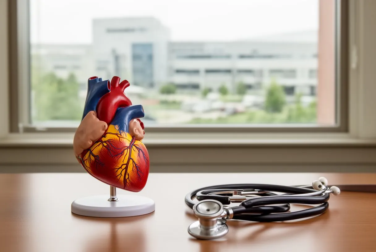
Why is heart surgery so different?
What makes heart surgery different from any other kind of operation — hernias, gallbladder, appendix, bone fractures, even neurosurgery — is the heart–lung machine we use, also known as the cardiopulmonary bypass machine. That single fact makes cardiac surgery a very different kind of intervention from all the others.
You will be wondering what this marvellous machine is. We will discuss how it works in a moment. First, it is important to know that before reaching heart surgery you will have gone through a relatively long process of visits to several specialists and, without doubt, many studies — echocardiogram, cardiac catheterisation, stress test, CT, MRI, laboratory tests, and we could go on listing several more.
After that, a group of expert specialists from different areas of cardiology, commonly known as the Heart Team, meet to discuss what the best treatment options might be, so that finally you and the people you love can make the decision that suits you best.
By this point you should already know the difference between an interventional cardiologist and a cardiac or cardiovascular surgeon.
Remember that most operations today are elective or planned, which is why this process can be carried out as part of the pre-surgical protocol. In emergencies the process is plainly different — rapid and expedited. For what to expect before, during and after the operation, see our guide to your heart surgery.
How heart surgery is performed: the details
Assuming the patient is already in the operating theatre, we will describe what is known as "open-heart" surgery, the approach most used worldwide. We will talk about minimally invasive procedures briefly afterwards.
The procedure is carried out under general anaesthesia — you will not remember it and will not be conscious — with several methods of advanced cardiovascular and neurological monitoring. For this, the anaesthetist will have placed various sensors and catheters in arteries and veins so the anaesthetic can be managed safely.
The cardiovascular anaesthetist has a very particular training and takes an active part in the procedure. The success of the surgery depends as much on them as on the surgeon. This is not a "normal" anaesthetist — it is a "super" anaesthetist.
Once that process is complete, the safety checklists are finished and we proceed with the surgery itself. It begins with an incision in the front of the chest, right in the middle. We then work deeper through the tissues until reaching the sternum, the bone you can feel in the middle of your chest. This is separated right down the middle. Don't worry — you will be under deep anaesthesia, feeling and remembering nothing.
Once the sternum is separated we have access to the structures surrounding the heart, and to the heart itself.
The heart is covered and protected by a sac we call the pericardium. This sac envelops the heart and holds a little fluid inside to prevent friction and wear, since the heart is a structure in constant motion.
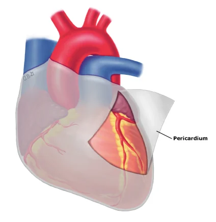
We open this sac at its front surface. It is at this moment that we can see the heart directly for the first time, beating, in all its splendour.
The structures visible here, besides the heart itself, are: the aorta, the main and largest artery of the body; the pulmonary artery, which carries deoxygenated blood to the lungs; the pulmonary veins, which return oxygenated blood to the heart; the superior and inferior vena cava, which bring blood from the whole body back to the heart; the innominate vein; and the aortic arch with the arteries leaving it towards the arms and the brain.
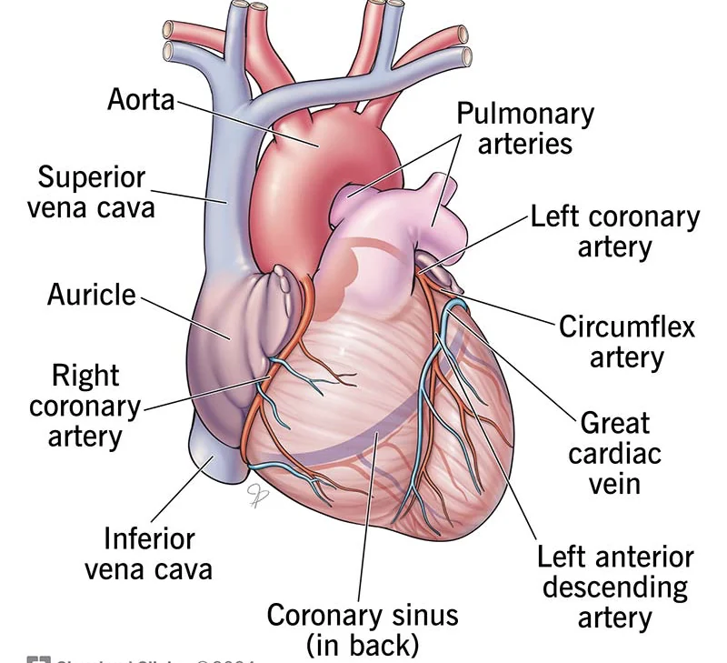
The next step is to prepare to connect this natural circulatory system to an artificial one — that is, to connect the patient to the heart–lung machine.
Connecting a patient to the cardiopulmonary bypass machine
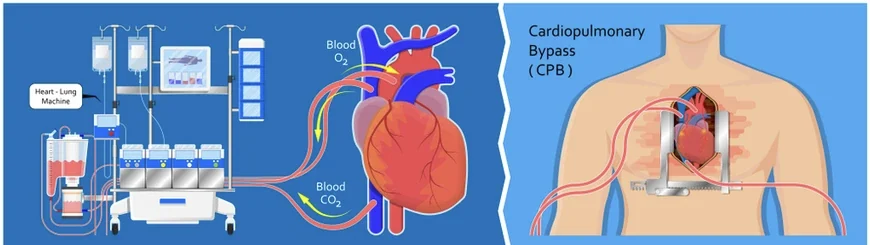
The cardiopulmonary bypass machine, also known as the extracorporeal circulation machine, is a crucial component in practically every open-heart operation. This device temporarily replaces the functions of the heart and lungs, allowing the surgeon to operate on a stopped or sleeping heart. The principle is that blood does not circulate through either the heart or the lungs while we are operating.
The process requires the participation of the whole team, like a great symphony.
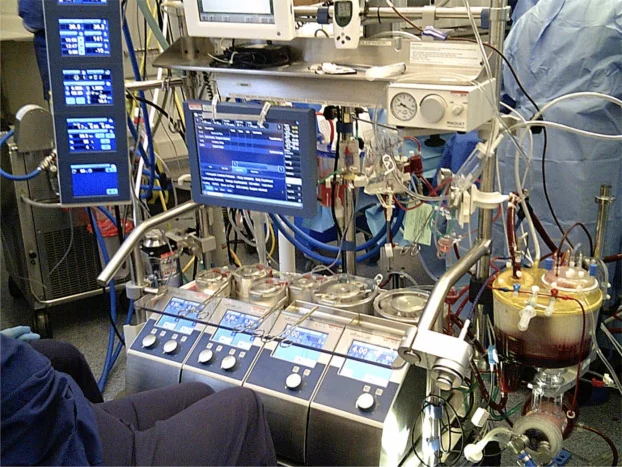
1. Connecting the cannulas
Connection to the bypass machine involves inserting cannulas into the great blood vessels to divert blood out of the body and then return it once oxygenated and filtered. Before this can begin, a very large dose of heparin is given to the patient to stop the blood clotting as it circulates through the artificial circuit.
- Venous cannula. A cannula is inserted into the right atrium, or into the superior and inferior vena cava, to divert deoxygenated blood away from the body and into the bypass machine before it enters the heart. This blood is carried through special tubing to the oxygenator — a kind of artificial lung.
- Arterial cannula. A cannula is inserted into the ascending aorta to return oxygenated blood to the patient's circulation. This blood is pumped from the bypass machine back into the body.
2. Starting the bypass machine
- Going on bypass. Once the cannulas are correctly placed and secured, the machine is switched on. The patient's deoxygenated blood flows into the machine, where it passes through an oxygenator that removes carbon dioxide and adds oxygen — as our own lungs normally would — and the oxygenated blood then returns through the special tubing to the cannula in the aorta, bypassing the circulation through the heart and lungs while maintaining adequate circulation to the rest of the body and organs such as the brain, kidneys and liver.
- Constant monitoring. The perfusionists, specialists in operating the machine, continuously monitor oxygen and carbon dioxide levels, blood pressure and blood flow, to be sure the blood returning to the patient is adequately oxygenated and at the correct temperature.
3. Maintaining body functions
- Oxygenation and circulation. During the operation the machine maintains oxygenation and blood circulation. The blood is cooled or warmed as needed to protect the organs and tissues.
- Anticoagulant medication. As mentioned, anticoagulants such as heparin are given to prevent the blood clotting while it is outside the body.
4. Performing the surgery
Once circulation on the bypass machine is adequate, a solution called cardioplegia is given, which reaches the coronary arteries. Cardioplegia has two functions: to put the heart to sleep, or stop it; and to protect it during this period without beating, providing the nutrients and the environment it needs so that it is not damaged and can beat again.
With the heart stopped and free of blood, the surgeon carries out the necessary procedure — a coronary bypass, a valve repair or replacement, the correction of a congenital heart defect, and so on.
Contrary to popular belief, the heart is not taken out of the body to be repaired. The surgery is done with the heart there in place. The only procedure where the heart is removed is a heart transplant.
The most common procedures performed in heart surgery are:
- Coronary revascularisation or bypass surgery
- Repair or replacement of the aortic valve
- Repair or replacement of the mitral or tricuspid valve
- Repair of congenital heart defects, present from birth
- Aortic aneurysms
- Aortic dissection
- Endocarditis
- Tumours of the heart
- Several others
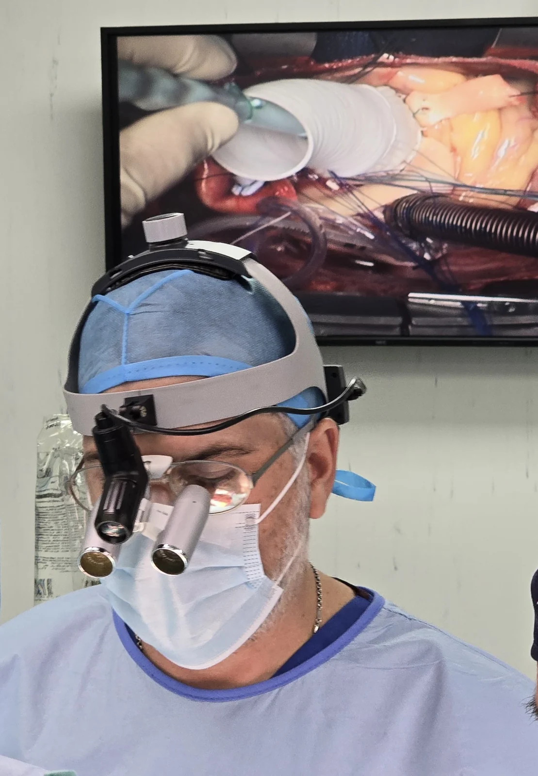
The length of the operation depends on the type of condition being treated. In the great majority of heart operations the bypass machine is used.
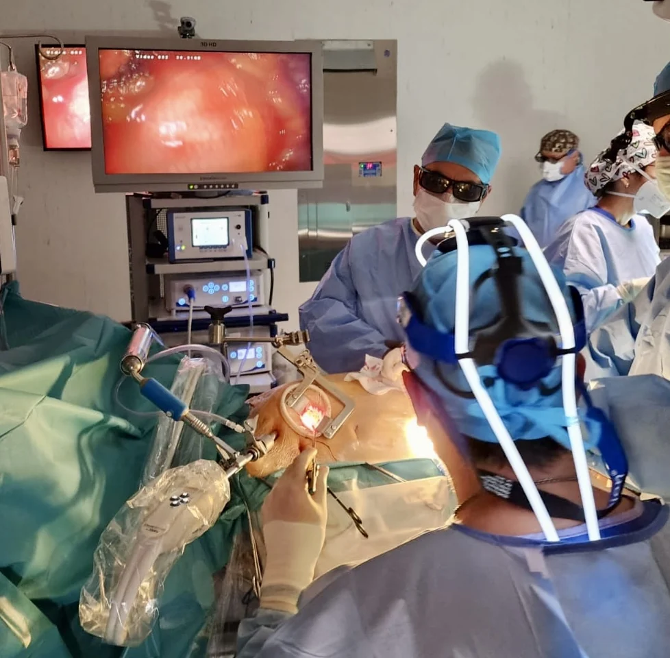
There are now minimally invasive cardiac techniques where we can avoid opening the sternum, using 3D cameras and special instruments. So it is not always necessary to perform "open-heart" surgery.
There are also new transcatheter valve replacement techniques with no surgical incision at all, carried out with special devices through the arteries. Most of the time these do not require the use of a bypass machine.
Age can vary a great deal depending on the condition. Heart surgery can be performed at practically any age — from a few hours old to patients over 80.
5. Coming off the bypass machine
- Restarting the heart. Once the surgery is complete, the patient's heart is restarted, letting their own blood enter the coronary arteries again and reperfuse the heart. This serves as a signal for it to "wake up" and take over its functions once more. Electric shocks are sometimes used to restore normal heart rhythm. Once we see that the patient's heart and lungs are able to resume their functions, the bypass machine gradually reduces its support until it ceases completely.
- Removing the cannulas. The venous and arterial cannulas are carefully withdrawn and the incisions in the blood vessels closed.
- Reversing the heparin. Once natural circulation is re-established and the cannulas removed, the effects of the heparin are reversed so the blood can clot again. The medicine used is called protamine.
- Closing. We check there are no sites of bleeding, and place special tubes to stop blood or inflammatory fluid accumulating inside the patient. Pacemaker wires are also placed on the heart. These are only temporary, in case the patient needs stimulation in the first hours after surgery. The sternum is closed with steel wires and the skin is sutured. Every patient goes out to the intensive care unit.
6. Postoperative monitoring
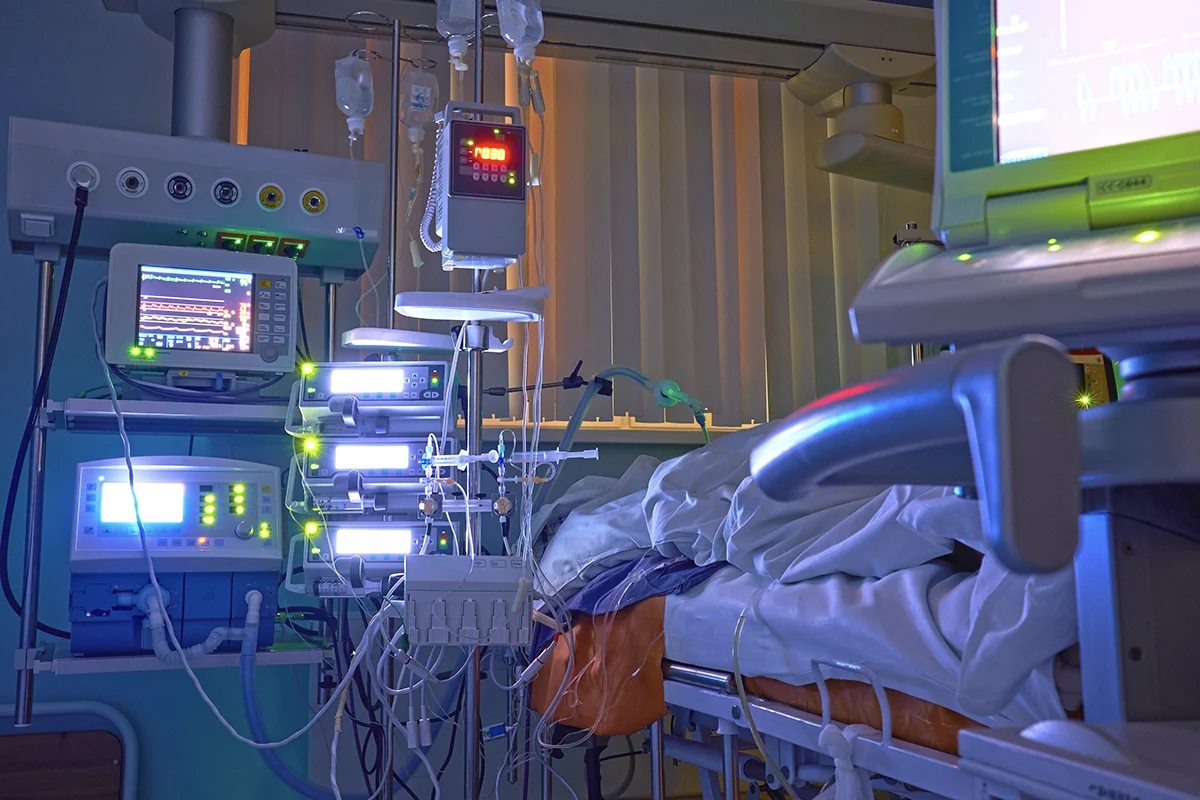
- Intensive care. After the operation the patient is transferred to the intensive care unit for close monitoring while recovering from the anaesthetic and the surgery.
- Recovery. The medical team continues to monitor vital signs, cardiac function and the healing of the wounds, making sure the patient receives the care and attention needed for a successful recovery.
An example: the heart bypass surgery process
Coronary bypass is the most common operation we perform. It is a good example of how heart surgery is performed in practice. The general steps are the ones described above. What changes is the repair itself.
- Harvesting the grafts. While the chest is being opened, we prepare the bypass conduits. The usual choice is the internal thoracic artery, from inside the chest wall. A vein from the leg or an artery from the forearm may also be used.
- Connecting below the blockage. With the heart stopped, each graft is sewn to a coronary artery beyond its narrowing. These vessels are only a few millimetres wide, so we use magnifying loupes and very fine sutures.
- Connecting the other end. Vein grafts are joined to the aorta, so blood flows from the aorta past the blockage. The internal thoracic artery usually stays attached at its origin.
- Checking the flow. Before closing, we make sure every graft fills well.
In selected patients the bypass can be done with the heart still beating, without the heart–lung machine. The rest of how heart surgery is performed stays much the same. Your surgeon will explain which approach suits you.
How long does each stage take?
Families waiting outside often ask why the operation seems so long. The repair itself is only part of the time. Anaesthesia, monitoring lines, opening the chest, connecting the machine, and careful closing all take time. For most planned operations the whole cardiac surgery procedure lasts around four to six hours. Complex cases or reoperations can take longer.
A longer operation is not a sign that something has gone wrong. Knowing how heart surgery is performed helps families understand the wait. Our team keeps them informed during the day.
Understanding what happens during heart surgery can ease a lot of fear. If you want to go through the heart surgery steps for your own operation, ask us at your consultation. Seeing how heart surgery is performed, one step at a time, often makes it feel far less mysterious.
A last word
Heart surgery is a wonderful thing. It is a privilege to have the chance to devote ourselves to it, and at the same time an enormous responsibility. Communication and a proper doctor–patient relationship are key to making the best decision about your treatment.
What makes this surgery so different from others, as we have said, is the artificial heart–lung machine. Connecting a patient to cardiopulmonary bypass is a meticulous and well-coordinated process that allows surgeons and the whole team to perform complex operations on the heart. This advanced procedure has saved countless lives and remains a fundamental part of modern cardiac surgery. With the support of an expert medical team and state-of-the-art technology, patients can face cardiac surgery with confidence and hope.
If you or a relative are facing a situation requiring heart surgery, remember that you are not alone. There are a great many people, trained and highly specialised, who can accompany you through this process.
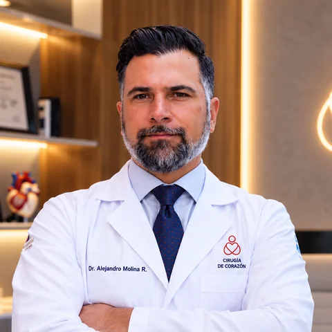
About the author
Dr Alejandro Molina is a cardiovascular surgeon practising in Guadalajara, Querétaro and Hermosillo. He trained in cardiothoracic surgery at the Universidad de Guadalajara, completed advanced training in cardiac surgery, minimally invasive surgery, robotic surgery and structural interventions at the University of Montreal, and a fellowship in structural cardiology and TAVR at CHU Bordeaux, France. He performed the first robotic mitral valve repair in the Ángeles Health System and the first robotic heart surgery in Guadalajara.
Medical degree: Universidad Autónoma de Guadalajara, Cédula Profesional 5757260 · Cardiothoracic surgery: Universidad de Guadalajara, Cédula de Especialidad 10768871 · Current certification, Consejo Nacional de Cirugía del Tórax, no. 447
Talk to a specialist
Request a free video consultation to clear up your questions and make a better-informed decision.





